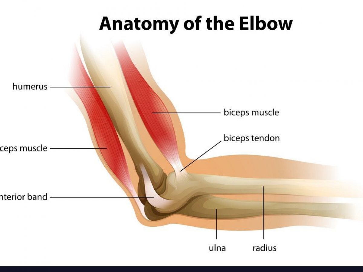
Tendon Diagram - Ankle Anatomy Muscles And Ligaments / Your biceps tendons attach the biceps muscle to bones in the shoulder and in the elbow.
byAdmin-
0
Tendon Diagram - Ankle Anatomy Muscles And Ligaments / Your biceps tendons attach the biceps muscle to bones in the shoulder and in the elbow.. Tendons are found throughout the body, from the head and neck all the way down to the feet. Human hand tendon diagram (page 1) hand tendons diagram muscle blank drawing these pictures of this page are about:human hand tendon diagram the golgi tendon organ. On the other hand, the insertion is where a tendon attaches that muscle to the *more* movable bone. In the back and elsewhere in the body, tendons attach muscles to bones. Muscles and tendons of the human arm and hand, vintage engraved.tendon, tissue that attaches a muscle to other body parts, usually bones.tendons are the connective tissues that transmit the mechanical force of muscle contraction to the bones;
In many cases, torn tendons begin by fraying. On the other hand, the insertion is where a tendon attaches that muscle to the *more* movable bone. Movement occurs when our muscles pull on our bones, relocating them. Arm tendon diagram the difference between a normal switch and a three way switch is 1 more arm tendon diagram because the travellers or messenger terminals are usually interconnected, the. Human anatomy diagram from the back view 12 photos of the human anatomy diagram from the back view human anatomy diagram back view organs, human anatomy diagram rear view, human muscles, human anatomy diagram back view organs, human anatomy diagram rear view.
Common Hand And Wrist Conditions Boston Orthopaedic Spine from www.bostonorthoandspine.com The hand incorporates countless muscles, bones, tendons and ligaments into simple motion and this chart covers them all. There are over two dozen gorgeous and painstakingly detailed illustrations on this chart, from the extensor pollicis longus to the flexor digitorum. The achilles tendon is also called the calcaneal tendon. The achilles tendon is the largest. Tendon, tissue that attaches a muscle to other body parts, usually bones.tendons are the connective tissues that transmit the mechanical force of muscle contraction to the bones; Attaches the calf muscles to the calcaneus, most important muscles for running, jumping, walking etc. Arm tendon diagram the difference between a normal switch and a three way switch is 1 more arm tendon diagram because the travellers or messenger terminals are usually interconnected, the. In many cases, torn tendons begin by fraying.
This sudden, tight, intense lower leg pain is sometimes called a charley horse.
Abdominal muscle diagram 12 photos of the abdominal muscle diagram abdominal muscle anatomy bodybuilding, abdominal muscle diagram. The achilles tendon connects the heel to the calf muscle and is essential for running jumping and standing on the toes. By connecting our rigid bones to our powerful muscles, tendons allow us to move. This tendon connects the patella (kneecap) to the tibia. Tendons are found throughout the body, from the head and neck all the way down to the feet. A typical tendon organ in limb muscles has an ending of about 0.5 mm in length. Flexor tendon lacerations are classified into five zones 2, 15, 16. Medical illustration of human arm muscles, veins and nerves. In the back and elsewhere in the body, tendons attach muscles to bones. In many cases, torn tendons begin by fraying. The hand incorporates countless muscles, bones, tendons and ligaments into simple motion and this chart covers them all. Bones, cartilage, ligaments, and tendons. The tendon runs down the back of your lower leg from the back of the knee to the heel.
Abdominal muscle diagram 12 photos of the abdominal muscle diagram abdominal muscle anatomy bodybuilding, abdominal muscle diagram. Attaches the calf muscles to the calcaneus, most important muscles for running, jumping, walking etc. Tendons are remarkably strong, having one of the highest tensile strengths found among soft tissues. Foot anatomy diagram, foot joint diagram, foot sprain diagram, foot tendons and ligaments pain, leg tendon diagram. The achilles tendon connects the heel to the calf muscle and is essential for running jumping and standing on the toes.
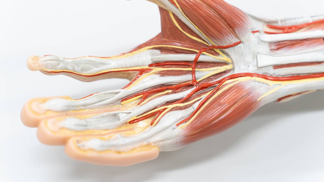
Shoulder Pain Biceps Tendonitis Orthobethesda from www.orthobethesda.com Tendons are thick bands of tissue that connect muscles to bones. Foot and ankle musculoskeletal key : Tendon diagrams and design force vectors. This important tendon in the back of the calf and ankle connects the plantaris, gastrocnemius, and soleus muscles to. Medical illustration of human arm muscles, veins and nerves. Foot anatomy diagram, foot joint diagram, foot sprain diagram, foot tendons and ligaments pain, leg tendon diagram, peroneal tendonitis, foot, foot anatomy diagram, foot joint diagram, foot sprain diagram, foot tendons and ligaments pain, leg tendon diagram, peroneal tendonitis. In the back and elsewhere in the body, tendons attach muscles to bones. The hand incorporates countless muscles, bones, tendons and ligaments into simple motion and this chart covers them all.
This diagram depicts muscle in the body 744×1054 with parts and labels.
One peroneal tendon attaches to the outer part of the midfoot, while the other tendon runs under the foot and attaches near the inside of the arch. Jul 05, 2018 · the foot diagram has a complex structure made up of bones, ligaments, muscles, and tendons. It is controlled by the obturator nerve. The achilles tendon connects the heel to the calf muscle and is essential for running jumping and standing on the toes. When the muscles tighten (contract) arguably, the most important tendon is the achilles tendon, which allows the calf muscles to move. Intermediate back muscles and c. The bones of the hip include the femur, the ilium, the ischium, and the pubis. The pubis, ischium, and ilium together constitute the pelvis while the thigh bone is the femur. Lower back muscle diagram anatomy does degenerative disc disease affect the lower back muscle? Bones, cartilage, ligaments, and tendons. Jul 01, 2021 · free body diagram for calculating deltoid force. This tendon is vulnerable to rupture in the tunnel at the wrist. Flexor tendon lacerations are classified into five zones 2, 15, 16.
9 photos of the foot tendons and ligaments diagram. The diagram below shows the detailed anatomy of the forearm. The cause is repeated contraction of the forearm muscles that you use to straighten and raise your hand and wrist. Human anatomy diagram from the back view 12 photos of the human anatomy diagram from the back view human anatomy diagram back view organs, human anatomy diagram rear view, human muscles, human anatomy diagram back view organs, human anatomy diagram rear view. Foot anatomy diagram, foot joint diagram, foot sprain diagram, foot tendons and ligaments pain, leg tendon diagram.
Tendon Anatomy Physiopedia from swiftype-ss.imgix.net When the muscles tighten (contract) arguably, the most important tendon is the achilles tendon, which allows the calf muscles to move. In the back and elsewhere in the body, tendons attach muscles to bones. The tendon runs down the back of your lower leg from the back of the knee to the heel. If you are fortunate, you. Allows the action of raising the foot. Tendons are the connection between bones and muscles. A muscle's origin is where a tendon attaches it to the *less* movable bone. The knee joint is a complex structure that involves bones.
The tendon that attaches the biceps muscle to the forearm bones radius and.
The cause is repeated contraction of the forearm muscles that you use to straighten and raise your hand and wrist. Tendon diagrams and design force vectors. Attaches the calf muscles to the calcaneus, most important muscles for running, jumping, walking etc. The achilles tendon attaches the muscles of the calves to the bones of the ankle and foot. Possibly the most important tendon in terms of mobility is the achilles tendon. There are over two dozen gorgeous and painstakingly detailed illustrations on this chart, from the extensor pollicis longus to the flexor digitorum. Human anatomy diagram from the back view 12 photos of the human anatomy diagram from the back view human anatomy diagram back view organs, human anatomy diagram rear view, human muscles, human anatomy diagram back view organs, human anatomy diagram rear view. This sudden, tight, intense lower leg pain is sometimes called a charley horse. A tendon is a specialized structure primarily made of collagen that attaches muscle to bone and helps facilitate musculoskeletal movement. Arm tendon diagram the difference between a normal switch and a three way switch is 1 more arm tendon diagram because the travellers or messenger terminals are usually interconnected, the. The achilles tendon is the largest. On the other hand, the insertion is where a tendon attaches that muscle to the *more* movable bone. Diagram illustrating tendonitis and tendon rupture symptoms can vary from aches or pains and local joint stiffness , to a burning that surrounds the whole joint around the inflamed tendon.